We design, fabricate, automate, and run platforms to address challenging questions in life sciences.
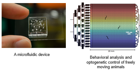
Microfluidic technology is at the heart of the platforms we build. Using a variety of techniques from soft lithography in cleanroom environment, to 3D printing or laser cutting, we can fabricate closed and open microfluidic networks, integrate actuators, modify surface properties, to create devices customized to the biological systems.
Moreover, we typically combine microfluidics with microscopy systems: from home-made scopes such as portable Raspberry Pi microscope or microfluidic-compatible light-sheet system to standard epifluorescence scope and commercial confocal microscope. Automation to control the hardware and actuate liquids, select imaging settings, and synchronize operations, plays an essential role to achieve high throughput. Finally, we develop software tools for analyzing and extract quantitative, high-content information from experimental data.
These advanced technological platforms allow us to investigate questions in neuroscience, cell biology, and behavior through a variety of biological systems: single cell, spheroids, organoids, Drosophila embryos, C. elegans embryos, C. elegans juvenile and adult animals.
Highlighted Lab Publications
- Lee, H. J., Vallier, J., & Lu, H. Microfluidic localized hydrogel polymerization enables simultaneous recording of neural activity and behavior in Caenorhabditis elegans. React. Chem. Eng. 9, 666-676 (2024). https://doi.org/10.1039/D3RE00516J
- Aubry, G., Milisavljevic, M., Lu, H., Automated and Dynamic Control of Chemical Content in Droplets for Scalable Screens of Small Animals. Small. 2022, 18. https://doi.org/10.1002/smll.202200319
- Le, K.N., Zhan, M., Cho, Y. et al. An automated platform to monitor long-term behavior and healthspan in Caenorhabditis elegans under precise environmental control. Commun Biol 3, 297 (2020). https://doi.org/10.1038/s42003-020-1013-2
- Cho, Y., Lee, S.A., Chew, Y.L., et al., Sensory Integration: Multimodal Stimulation in a Microfluidic Device Facilitates Studies of Interneurons in Sensory Integration in C. elegans. Small. 2020, 16. https://doi.org/10.1002/smll.201905852
- Cho, Y., Porto, D., Hwang, H., et al., Automated and controlled mechanical stimulation and functional imaging in vivo in C. elegans. Lab Chip. 2017, 17. https://doi.org/10.1039/C7LC00465F
- Lee, H., Kim, S.A., Coakley, S., et al., A multi-channel device for high-density target-selective stimulation and long-term monitoring of cells and subcellular features in C. elegans. Lab Chip. 2014, 23. https://doi.org/10.1039/C4LC00789A
- Chung, K., Rivet, C., Kemp, M., and Lu, H., Imaging Single-Cell Signaling Dynamics with a Deterministic High-Density Single-Cell Trap Array. Analytical Chemistry. 2011, 83. https://doi.org/10.1021/ac2011153
- Chung, K., Kim, Y., Kanodia, J. et al., A microfluidic array for large-scale ordering and orientation of embryos. Nat Methods. 2011, 8. https://doi.org/10.1038/nmeth.1548
- Chung K., Crane M.M., Lu H., Automated on-chip rapid microscopy, phenotyping and sorting of C. elegans. Nat Methods. 2008, 5. https://doi.org/10.1038/nmeth.1227