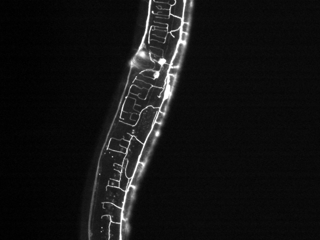
We develop integrated systems combining advanced fluorescent imaging techniques, such as light sheet microscopy with systems for stimulating and manipulating organisms, including microfluidic platforms for mechano-, thermo- and chemo-stimulation, and optogenetic manipulation of targeted neurons and muscles for freely moving worms. This allows us to assess the direct effect of environmental stimuli on single targeted neurons, groups of neurons, and the whole brain.
Highlighted Lab Publications
- Chaudhary, S., Lee, S.A., Li, Y., Patel, D., Lu, H., Graphical-model framework for automated annotation of cell identities in dense cellular images. eLife. 2021. https://doi.org/10.7554/eLife.60321
- Porto, D., Matsunaga, Y., Franke, B., et al., Conformational changes in twitchin kinase in vivo revealed by FRET imaging of freely moving C. elegans. eLife 2021. https://doi.org/10.7554/eLife.66862
- Porto, D.A., Giblin, J., Zhao, Y. et al., Reverse-Correlation Analysis of the Mechanosensation Circuit and Behavior in C. elegans Reveals Temporal and Spatial Encoding. Sci Rep 2019, 9. https://doi.org/10.1038/s41598-019-41349-0
- Stirman, J., Crane, M., Husson, S. et al., A multispectral optical illumination system with precise spatiotemporal control for the manipulation of optogenetic reagents. Nat Protoc. 2012, 7. https://doi.org/10.1038/nprot.2011.433
- Stirman, J., Crane, M., Husson, S. et al., Real-time multimodal optical control of neurons and muscles in freely behaving Caenorhabditis elegans. Nat Methods. 2011, 8. https://doi.org/10.1038/nmeth.1555
